RODOLFO C. PUCHE
Laboratorio de Biología Ósea, Facultad de Ciencias Médicas, Universidad Nacional de Rosario, Santa Fe, Argentina
Resumen Determinantes genéticos de hipovitaminosis D. La hipovitaminosis D, definida por bajos niveles
séricos de 25(OH)D (<12 ng/ml), es un reconocido problema de salud pública mundial. La deficiencia de vitamina D a largo plazo resulta en una disminución de la mi neralización ósea, hiperparatiroidismo secundario, pérdida de hueso cortical (patogénesis de la osteoporosis y fracturas de cadera), diferenciación y división de varios tipos de células, fuerza muscular, diabetes tipo 2, pres ión arterial, etc. Estudios genéticos han demostrado que algunos “polimorfismos de un solo nucleótido” (SNP) están relacionados con bajas concentraciones séricas de 25(OH)D a través de reducción en la actividad de las enzimas implicadas en la síntesis de 1α,25(OH)2D. Los médicos no necesitan indicar un estudio genético para identificar a la insuficiencia de vitamina D de causa genética. Bastará con instruir a los pacientes sobre su propio cuidado y controlar la ingesta de vitamina D y los niveles séricos de 25(OH)D hasta que estos últimos sean adecuados. En general, la literatura revela que las consecuencias de la hipovitaminosis D sobre la salud ósea se observan en las personas añosas y con poca
frecuencia en sujetos jóvenes. Una explicación probable para esta situación es: si la tasa de remodelación ósea lo permite, el tejido óseo tiene factores endógenos (genéticos, hormonales) y exógenos (dieta, actividad física) que pueden compensar las variables de la salud ósea. Las consecuencias del déficit de vitamina D sobre la salud ósea aún no se conocen completamente.
Palabras clave: hipovitaminosis D, polimorfismos de un solo nucléotido, remodelación ósea, edad
Abstract Hypovitaminosis D, defined by low serum levels of 25(OH)D, is a recognized worldwide public health
problem. The most accepted definition considers that deficiency occurs with serum levels fall below 12 ng/ml of 25(OH)D. Long term vitamin D deficiency results in decreased bone mineralization, secondary hyperparathyroidism, increased cortical bone loss (pathogenesis of osteoporosis and hip fractures), differentiation and division of various cell types, muscle strength, diabetes type 2, blood pressure, etc. Twin- and family-based studies indicate that genetic factors influence serum 25(OH)D levels. Genetic studies have shown single-nucleotide polymorphisms (SNPs) are linked to low serum 25(OH)D concentrations through changes in the activity of the enzymes of the 1α,25(OH)2D metabolic pathway. Carriers of high genetic risk scores would need a h igher amount of vitamin D supplementation to achieve adequate serum 25(OH)D concentrations. Clinicians would not need to indicate studies to identify patients with vitamin D insufficiency of genetic origin. They should instruct their patients on their own care, to control the intake of vitamin D and the serum 25(OH)D levels until the latter are adequate.
Overall, the literature reveals that the consequences of hypovitaminosis D on bone health are observed in old and infrequently in young subjects. A probable explanation for the latter is: if the rate of bone remodeling allows it, bone tissue has endogenous (genetics, hormones) and exogenous determinants (diet, physical activity) that may compensate the variables of bone health. The consequences of vitamin D deficit on bone health, has not been completely uncovered.
Key words: hypovitaminosis D, single-nucleotide-polymorphisms, genes, bone remodeling, age
Dirección postal: Rodolfo C. Puche, Laboratorio de Biología Ósea, Facultad de Ciencias Médicas, UNR, Santa Fe 3100, 2000 Rosario, Santa Fe, Argentina
e-mail: rodolfopuche@gmail.com
Hypovitaminosis D, defined by the low serum levels of 25(OH)D, is a worldwide public health problem 1. The definitions of the nutritional status of vitamin D have varied in recent years. At present, the most accepted definition is that that considers vitamin D3 deficiency when serum 25(OH)D concentration is below 12 ng/ml 2. Vitamin D plays a primary physiological role in maintaining extracellular calcium ion levels indispensable for the functioning of many metabolic processes and neuromuscular activity. The vitamin influences calcium levels primarily by controlling the absorption of calcium from the intestine, through direct effects on bone and through its effects on parathyroid hormone (PTH) secretion 3,4.
Long term vitamin D deficiency results in decreased bone mineralization, secondary hyperparathyroidism, and increased cortical bone loss. These factors that have been associated to the pathogenesis of osteoporosis and hip fractures 5, 6. Vitamin D3 yield the biologically active 1a, 25(OH)2D, which acts through specific vitamin D receptors to regulate not only calcium metabolism, but also differentiation and division of various cell types 7, 8. It has been reported that in addition to its pivotal role in calcium metabolism and bone mineralization, vitamin D may play a role in muscle strength 9, 10, diabetes type 2 11, blood pressure 12, etc.
Glossary
Allele: A variant form of a given gene. Sometimes, the presence of different alleles of the same gene can result in different observable phenotypic traits, such as different skin pigmentation. The addition of the letter A (adenine), C (cytosine) or T (thymine) before the term “allele”, indicates the different nucleotide.
SNPs: Single nucleotide polymorphisms (SNPs), are the most common type of genetic variation. For example, in a determined SNP the nucleotide cytosine (C) may be replaced with the nucleotide thymine (T), in a certain stretch of DNA. SNPs occur normally throughout a person’s DNA.
They occur almost once in every 1,000 nucleotides on average, which means there are roughly 4 to 5 million SNPs in a person’s genome. These variations may be unique or occur in many individuals; scientists have found more than 100 million SNPs in populations around the world. Most commonly, these variations are found in the DNA between genes. They can act as biological markers, helping scientists locate genes that are associated with disease. Most SNPs have no effect on health or development. Some of these genetic differences, however, because they affect the enzymes of the metabolic pathway of vitamin D, have proven to be important in the determination of vitamin D deficiency.
Genes associated with the synthesis and physiological effects of vitamin D3
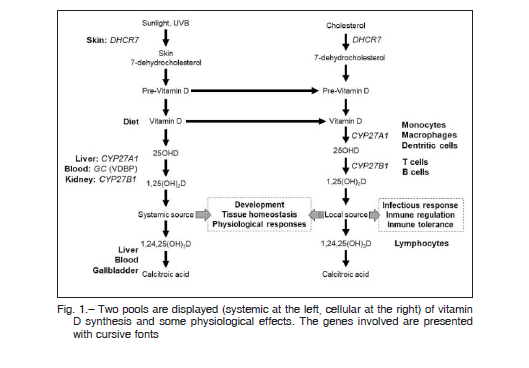
The Figure 1 displays two pools (systemic at the left, cellular at the right) of vitamin D3 synthesis and some physiological effects. The genes involved are presented with cursive fonts. Their products (enzymes) are described below.
DHCR7
The DHCR7 gene encodes instructions for making the enzyme 7-dehydrocholesterol reductase, responsible for the final step in cholesterol production in many types of cells. Specifically, this enzyme converts 7-dehydrocholesterol to cholesterol13.
CYP27R1
Cheng et al 14 reported that this gene encodes vitamin D 25-hydroxylase. This enzyme is a member of the cytochrome P450 superfamily. The cytochrome P450 proteins are monooxygenases which catalyze many reactions involved in drug metabolism and synthesis of cholesterol, steroids and other lipids. The protein encoded by this gene localizes to the inner mitochondrial membrane where it hydroxylates 25-hydroxyvitamin D at the 1a position. This reaction synthesizes 1a, 25-(OH)2D, the active form of vitamin D3, which binds to the vitamin D receptor and regulates calcium metabolism. Thus this enzyme regulates the level of biologically active vitamin D and plays an important role in calcium homeostasis. Mutations in this gene can result in vitamin D-dependent rickets type I.

GC
The Gc protein 15 (human group-specific component), is a well-known vitamin D-binding protein (VDBP) or Gc globulin, a 55 kDa serum protein secreted by the liver and belonging to the albumin superfamily. It has physiological functions that include involvement in vitamin D transport and storage, scavenging of extracellular G-actin, enhancement of the chemotactic activity of C5 for neutrophils in inflammation and macrophage activation.
CYP27B1
This gene 13 encodes a member of the cytochrome P450 superfamily of enzymes: 25(OH)D-1-alpha-hydroxylase. This enzyme is located in the proximal tubules of the kidney and a variety of other tissues, including skin, immune cells, and bone.
Reports associating genes with low levels of 25(OH)D
In northern latitudes (= 40° North), low vitamin D status in humans, measured as 25(OH)D concentrations, is common during winter months. This is because vitamin D cannot be synthesized in the skin due to the lack of solar ultraviolet B radiation (UVB) and because the average dietary intake of vitamin D is insufficient 16. Twin- and family-based studies have shown that genetic factors may influence 25(OH)D concentrations 17, 18 Several candidate gene studies have shown single-nucleotide polymorphisms (SNPs) to influence 25(OH)D concentrations 19-24. These SNPs are located in the group-specific component also known as Gc globulin (GC) and in or near genes involved in vitamin D synthesis, activation or degradation. These findings indicate that 25(OH)D concentrations do not only depend on vitamin D intake and sun exposure, but also that genetic factors may help to identify individuals at risk of low vitamin D status. This paper reviews the genome-wide association studies (GWAS) of Wang et al25 and Nissen et al 26.
Summary of Wang et al and Nissen et al reports 25, 26
These two reports produced complementary information. The Wang´s report contributed to elucidate the architecture of vitamin D insufficiency providing a better understanding of the regulation of the vitamin metabolism and identifying genetic variants useful for the identification of individuals at substantially elevated risk for vitamin D insufficiency 25. The Nissen´s report assessed the effects of real-life use of vitamin D3-fortified bread and milk on 25(OH)D serum concentrations in relation to common genetic variants of 25-hydroxylase and vitamin D binding protein (GC) 26.
Studies design
Wang´s studies were conducted only with white individuals of European descent. They measured plasma 25(OH) D levels and performed the genome analysis on 16,125 individuals of European descent drawn from five epidemiological cohorts, plus five additional cohorts (n = 9,366) with genome-wide association data 25. The Nissen´s study was a double blinded, randomized placebo-controlled intervention trial performed with apparently healthy Danish children and adults. A total of 201 Danish families with dependent children, 4-60 years of age, randomly drawn from the Danish Civil Registration System, participated in the study. The study ensured a large age span and one weakness: some of the variables associated with 25(OH)D serum concentrations were quantified by self-reported questionnaire data. Families were randomly allocated to either vitamin D3-fortified bread and milk or non-fortified placebo bread and milk during a 6-month winter period without sunlight exposure. At the end of the study, a total of 758 participants (no fortification group: n = 384; fortification group: n = 384) had complete questionnaire data, genotypes and 25(OH)D concentrations measured 26.
Variables investigated
The selected SNPs for replication had meta-analytic Pvalues < 5 × 10⁻⁸ in the discovery samples. For further analysis, the selected SNPs were located at or near six pre-specified vitamin D pathway candidate genes: vitamin D receptor (VDR), 1-a-hydroxylase (CYP27B1), 25-hydroxylase (CYP2R1), 24-hydroxylase (CYP24A1), vitamin D binding protein (GC, VDBP), and 27- and 25-hydroxylase (CYP27A1). The selected SNPs were assessed for 25(OH)D association in the de novo replication samples, and were combined to produce P-values across these 15 studies 27.
Nissen el al previously found that two SNPs in 25-hydroxylase and another two in vitamin D binding protein, predicted baseline 25(OH)D concentrations 28. None of the four SNPs were in linked disequilibrium with each other, which means that they are not in random association of alleles at different loci in a given population. The main focus of this study was set on the influence of these four SNPs on 25(OH)D concentrations in participants allocated to either vitamin D-fortified bread and milk or non-fortified bread and milk during winter 27.
Results
The data obtained in Wangs´ study established a role for common genetic variants in the regulation of circulating 25(OH)D levels 25. Indeed, the presence of identified alleles at the confirmed loci improve the understanding of vitamin D homeostasis and may assist in the identification of a subgroup of Caucasians at risk for vitamin D insufficiency of genetic origin. The existence of these loci in a given subject more than double the odds of vitamin D insufficiency occurrence. Measurements of serum 25(OH) D concentrations were conducted by isotope dilution liquid chromatography tandem mass spectrometry (LC–MS/ MS). The cut-off value of 25(OH)D > 20 µg/ml defined the requirement for optimal bone health for the majority of the population, and the cut-off value <12 ng/ml defined the 25(OH)D concentration at which adverse effects on bone health may be expected 30.
The contribution of vitamin D from intakes of vitamin D3-fortified bread and milk was calculated based on the self-reported consumption frequencies, amount and the measured vitamin D3 contents in the fortified products (5.2 ± 0.3 µg/100 g in wheat bread, 4.3 ± 0.3 µg/100 g in rye bread and 0.38 µg/100 ml in milk).
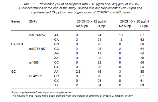
Nissen et al. demonstrated that carriers with accumulated risk alleles (high genetic risk score, GRS) of the SNPs of 25-hydroxylase and vitamin D binding protein are more prone to be vitamin D deficient compared to carriers of a low GRS (Table 1) 26. Carriers of high GRS require a greater amount of vitamin D supplementation to achieve adequate 25(OH)D serum levels. The results strongly indicated that clinicians would not need to indicate a genetic study to their patients with vitamin D insufficiency. They will have to instruct their patients and control the intake of vitamin D and the serum 25(OH)D levels until the latter are adequate.
Vitamin D safety
Serum concentrations of 25(OH)D up to 100 ng/ml are regarded safe in the general population of children and adults, although in preterm neonates (a specific group), an increased risk of hypercalcemia has been reported at the 25(OH)D3 values > 80 ng/ml 31. Hypercalcemia and hypercalciuria may occur when vitamin D intake is uncontrolled resulting in levels above 150-200 ng/ml2. According to reviewers of this subject, exceptional conditions comprise individuals with vitamin D hypersensitivity, and also with idiopathic infantile hypercalcemia 33,34, Williams-Beuren syndrome 35, granulomatous diseases 36 and some lymphomas. No evidence exists until now that these values may be exceeded when appropriate doses of vitamin D are used.
Hypovitaminosis D in Argentina
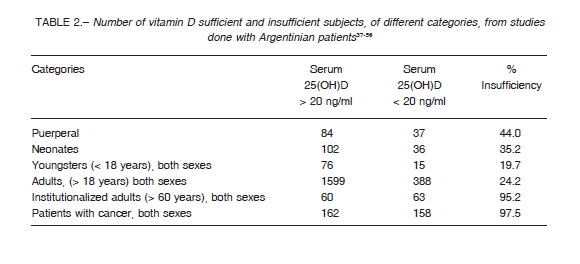
The serum levels of 25(OH)D of assumedly healthy inhabitants of Argentina have been the matter of additional nineteen reports, published since 1987 37-56. Only thirteen of those papers reported the number of subjects with insufficiency: a total of 2337 assumedly healthy persons, 476 of which (20.4%) were in insufficiency. The incidence would have been higher if the figures of institutionalized adults > 60 years or age and patients with cancer (nearly all of which were in insufficiency) were included (Table 2).

The report by Duran et al. on the Encuesta Nacional de Nutrición y Salud (2004-2005) describe the nutritional status of inhabitants under 5 years of age for our country as a whole and by regions 57. In disagreement with the data of Table 2, the report states that vitamin D deficiency in children aged 6-23 months of age, in Patagonia, was 2.8% (the report states that the data is based in actual measurements of serum 25(OH)D but no supporting data were included). It can be concluded that the deficiency of vitamin D is a definite health problem in Argentina. It would be desirable to have national surveys of similar quality as those existing in other countries like, The Danish National Health Service Register 58, or The US National Health and Nutrition Examination Survey 59.
The effects of vitamin D deficit on bone health
The consequences of hypovitaminosis D are easily observed in children on account of their high rate of bone remodeling. In adults, the consequences of the deficit can be observed (without the aid of invasive measures) when the deficit has been chronic for many years. This observation poses the question: “why bone disease is not easily evident in short-term vitamin D deficiency in adults? An answer, probably adequate, could be: if the rate of bone remodeling allows it, bone tissue has endogenous (genetic, hormones) and exogenous determinants (diet, physical activity) that may compensate each other. The rate of bone remodeling, assessed by stature, decays continuously (with the exception of the pubertal spurt) along lifespan 60.
The vitamin status, assessed by serum 25(OH)D, of young adults and the elderly has been reviewed by McKenna from 1971 to 1990 61. Hypovitaminosis D and related abnormalities in bone chemistry are reported in all elderly populations. The vitamin D status in young adults and the elderly varies widely with the country of residence (in McKenna´s report, studies were grouped according to geographic regions: North America; Scandinavia; Central and Western Europe). Adequate exposure to summer sunlight is the essential mean to obtain vitamin D, but oral intake augmented by both fortification and supplementation of foods are necessary to maintain baseline stores. McKenna states that it seems likely that the elderly would benefit additionally from a daily supplement of 10 micrograms of vitamin D and insist in that all countries should adopt a food fortification policy. Literature reports, however, indicate that vitamin D supplementation is not the only requirement to insure bone health.
Oliveri et al carried out a study to evaluate the possible influence of chronic winter vitamin D deficiency and higher winter parathyroid hormone (PTH) levels on bone mass in prepuberal children and young adults 62. The study was carried out in male and female Caucasian subjects, recruited from two cities of Argentina: Ushuaia (55° South), where the population is known to have low winter 25(OH)D levels and higher levels of PTH in winter than in summer, and Buenos Aires (34° South), where ultraviolet radiation and vitamin D nutritional status in the population are adequate all year round 45. A total of 163 prepuberal children (8.9 ± 0.7 years) and 234 young adults (22.9 ± 3.6 years) who had never received vitamin D supplementation were evaluated. Similar results were obtained in age-sex matched groups in bone mineral content (BMC) and bone mineral density (BMD) of the ultradistal and distal radius. In conclusion, peripheral BMD and BMC were similar in children and young adults from Ushuaia and Buenos Aires in spite of the previously documented difference between both areas regarding ultraviolet radiation and winter vitamin D status.
Callegari et al explored determinants of bone parameters in 326 young women, 16-25 years of age 63. Serum 25(OH)D was measured and bone health was assessed using dual-energy X-ray absorptiometry and peripheral quantitative computed tomography. Mean (± SD) serum 25(OH)D was 28 ± 11 ng/ml and the prevalence of vitamin D deficiency (serum 25(OH)D < 20 ng/ml) was 26%. Serum 25(OH)D levels were not associated with the two bone health parameters evaluated. Most bone parameters were found positively associated with height and lean mass.

Shah et al aimed to determine whether bone fragility was present in 150 subjects, excluding persons aged younger than 20 years and patients with hypercalcemia (serum calcium > 2.6 mmol/l), and chronic kidney disease (glomerular filtration rate < 60 ml/min per 1.73 m2)64. Bone health was assessed measuring serum 25(OH)D, serum C-terminal telopeptide of type 1 collagen, serum procollagen type 1 N-terminal propeptide, low areal bone mineral density (aBMD), distal radius microstructure deterioration and reduced matrix mineralization density (MMD). No associations were detected between serum 25(OH)D levels, aBMD, trabecular density, cortical porosity, or MMD.
On a sample of 1236 women aged = 50 years in the baseline survey, Tamaki et al. collected information regarding fractures during a 15-year follow-up period 65. The analysis included 1211 women without early menopause or diseases affecting bone metabolism. Over 15 years of follow up, 269 clinical (224 non-vertebral, 149 fragility) fracture events were confirmed. Incidence rates categorized by 25(OH)D levels (<10, 10-20, 20-30, and = 30 ng/ml) indicated a significant divergence for any clinical fractures in 5 years and for non-vertebral fractures in 5, 10, and 15 years.
It is well-established that prolonged and severe vitamin D deficiency leads to osteomalacia in adults. Sub-optimal vitamin D status has been reported in many populations but it is a particular concern in older people. Over extended periods of time, insufficiency has been associated with increased bone loss and secondary hyperparathyroidism leading to increased fracture risk 66. Sufficiency has been regarded as the point at which further intakes will have no additional beneficial effects on PTH and calcium metabolism in regard to bone health. However, the cutoff values for sufficiency are still under debate. A study has demonstrated concentrations of > 30 ng/ml or higher, 40-80 ng/ml, as optimal 67. However, the formation of vitamin D from sunlight is a self-limiting reaction; thus preventing toxicity from sun exposure. The development of bone disease in later life is related to the attainment of maximum peak bone mass and the maintenance of bone mass in adulthood 68.
The story of bone health as a consequence of vitamin D deficit is not, yet, completely uncovered 69. Research has shown that inadequate vitamin D intakes over long periods of time can lead to bone demineralization. These alterations result in the up-regulation of some components of the immune system as well as diminished function of others 70, 71. Changes include decreased mature lymphocyte function, decreased replication of hematopoietic cells and an up-regulation in the production of pro-inflammatory cytokines such as interleukin 6 and tumor necrosis factoralpha These pro-inflammatory cytokines have been associated with increased bone metabolism and osteoporosis is often considered to be an inflammatory condition. The immunoregulatory mechanisms of vitamin D may thus modulate the effect of these cytokines on bone health and subsequent fracture risk.
Conflict of interests: None to declare
References
1. Mithal A, Wahl DA, Bonjour J, et al. Global vitamin D status and determinants of hypovitaminosis D. Osteoporos Int 2009; 20: 1807-20.
2. Ross A, Manson J, Abrams S, et al. The 2011 report on dietary reference intakes for calcium and vitamin D from the Institute of Medicine: What clinicians need to know. J Clin Endocrinol Metab 2011; 96: 53-8.
3. Parfitt AM, Gallagher JC, Heaney RP, et al. Vitamin D and bone health in the elderly. Am J Clin Nutr 1982; 36: 1014-31.
4. Holick M. Vitamin D: the underappreciated D-lightful hormone that is important for skeletal and cellular health. Curr Opin Endocrinol Diabetes Obes 2002; 9: 87-98.
5. Rizzoli R, Bonjour J. Dietary protein and bone health. J Bone Miner Res 2004; 19: 527-31.
6. Lips P. Vitamin D deficiency and secondary hyperparathyroidism in the elderly: consequences for bone loss and fractures and therapeutic implications. Endocr Rev 2001; 22: 477-501.
7. Gniadecki R. Effects of 1,25-dihydroxyvitamin D3 and its 20-epi analogues (MC 1288, MC 1301, KH 1060), on clonal keratinocyte growth: evidence for differentiation of keratinocyte stem cells and analysis of the modulatory effects of cytokines. Br J Pharmacol 1997; 120: 1119-27.
8. Zhao X, Feldman D. Regulation of vitamin D receptor abundance and responsiveness during differentiation of HT-29 human colon cancer cells. Endocrinology 1993; 132: 1808-14.
9. Bischoff HA, Stahelin HB, Urscheler N, et al. Muscle strength in the elderly: its relation to vitamin D metabolites. Arch Phys Med Rehabil 1994; 80: 54-58.
10. Holick MF. Vitamin D and the skin: photobiology, physiology and therapeutic efficacy for psoriasis. In: Heersche JNM, Kanis JA (eds), Bone and mineral research. Amsterdam: Elsevier, 1990, p 313-66.
11. Pittas AG, Harris SS, Stark PC, et al. The effects of calcium and vitamin D supplementation on blood glucose and markers of inflammation in nondiabetic adults. Diabetes Care 2007; 30: 980-6.
12. Rostand SG. Ultraviolet light may contribute to geographic and racial blood pressure differences. Hypertension 1979; 30: 150-6.
13. Genetics Home Reference NIH. In: https://ghr.nlm.nih.gov/gene/DHCR7; accessed January 2019.
14. Cheng JB, Levine M, Bell N, et al. Genetic evidence that the human CYP2R1 enzyme is a key vitamin D 25-hydroxylase. Proc Natl Acad Sci USA 2004; 101: 7711-5.
15. Nagasawa H, Uto Y, Sasaki H, et al. Gc Protein (Vitamin D-binding Protein): Gc Genotyping and GcMAF Precursor Activity. Anticancer Res 2005; 25: 3689-96.
16. Thuesen B, Husemoen L, Fenger M, et al. Determinants of vitamin D status in a general population of Danish adults. Bone 2012; 50: 605-10.
17. Engelman CD, Fingerlin TE, Langefeld CD, et al. Genetic and environmental determinants of 25-hydroxyvitamin D and 1,25-dihydroxyvitamin D levels in Hispanic and African Americans. J Clin Endocrinol Metab 2008; 93: 3381-8.
18. Karohl C, Su S, Kumari M, et al. Heritability and seasonal variability of vitamin D concentrations. Am J Clin Nutr 2010; 25: 1393-8.
19. Sinotte M, Diorio C, Berube S, et al. Genetic polymorphisms of the vitamin D binding protein and plasma concentrations of 25-hydroxyvitamin D in premenopausal women. Am J Clin Nutr 2009; 25: 634-40.
20. Shea MK, Benjamin EJ, Dupuis J, et al. Genetic and nongenetic correlates of vitamins K and D. Eur J Clin Nutr 2009; 63: 458-64
21. Bu FX, Armas L, Lappe J, et al. Comprehensive association analysis of nine candidate genes with serum 25-hydroxy vitamin D levels among healthy Caucasian subjects. Hum Genet 2010; 128: 549-56.
22. Ahn J, Yu K, Stolzenberg-Solomon R, et al. Genome-wide association study of circulating vitamin D levels. Hum Mol Genet 2010; 19: 2739-45.
23. Zhang Z, He JW, Fu WZ, et al. An analysis of the association between the vitamin D pathway and serum 25-hydroxyvitamin D levels in a healthy Chinese population. J Bone Miner Res 2013; 28: 1784-92.
24. Engelman CD, Meyers KJ, Iyengar SK, et al. Vitamin D intake and season modify the effects of the GC and CYP2R1 genes on 25-hydroxyvitamin D concentrations. J Nutr 2013; 25: 17-26.
25. Wang TJ, Zhang F, Brent Richards J, et al. Common genetic determinants of vitamin D insufficiency: a genomewide association study. Lancet 2010; 376: 180-8.
26. Nissen J, Vogel U, Raven-Haren G, et al. Real-life use of vitamin D3 fortified bread and milk during winter season: the effects of CYP2R1 and GC genes on 25-hydroxyvitamin D concentrations in Danish families, the VitmaD study. Genes Nutr 2014; 9: 413-29.
27. Skol AD, Scott LJ, Abecasis GR, Boehnke M. Joint analysis is more efficient than replication-based analysis for two-stage genome-wide association studies. Nat Genet 2006; 38: 209-13.
28. Nissen J, Rasmussen LB, Ravn-Haren G, et al. Common variants in CYP2R1 and GC genes predict vitamin D Concentrations in healthy Danish children and adults. PLos One 2014; 9: e89907.
29. Gozdzik A, Zhu J, Wong BY, et al. Association of vitamin D binding protein (VDBP) polymorphisms and serum 25(OH)D concentrations in a sample of young Canadian adults of different ancestry. J Steroid Biochem Mol Biol 2011; 127: 405-12.
30. Ross A, Manson J, Abrams S, et al. The 2011 report on dietary reference intakes for calcium and vitamin D from the Institute of Medicine: what clinicians need to know. J Clin Endocrinol Metab 2011; 96: 53-8.
31. Czech-Kowalska J, Dobrzanska A, Pleskaczynska A, et al. Vitamin D status in premature infants at term. Bone 2009; 45 (Suppl 2): S107.
32. Lukaszkiewicz J, Prószynska K, Lorenc RS, et al. Hepatic microsomal enzyme induction: treatment of vitamin D poisoning in a 7 month old baby. Br Med J (Clin Res Ed) 1987; 295: 1173.
33. Jones G, Kottler ML, Schlingmann KP. Genetic diseases of vitamin D metabolizing enzymes. Endocrinol Metab Clin North Am 2017; 46: 1095-117.
34. Pronicka E, Ciara E, Halat P, et al. Biallelic mutations in CYP24A1 or SLC34A1 as a cause of infantile idiopathic hypercalcemia (IIH) with vitamin D hypersensitivity: molecular study of 11 historical IIH cases. J Appl Genet 2017; 58: 349-53.
35. Lameris AL, Geesing CL, Hoenderop JG, Schreuder MF. Importance of dietary calcium and vitamin D in the treatment of hypercalcaemia in Williams-Beuren syndrome. J Pediatr Endocrinol Metab 2014; 27: 757-61.
36. Bosch X. Hypercalcemia due to endogenous overproduction of active vitamin D in identical twins with cat-scratch disease. JAMA 1998; 279: 532-4.
37. Ladizesky M, Oliveri B, Mautalen C. Niveles séricos de 25-hidroxi-vitamina D en la población normal de Buenos Aires. Medicina (B Aires) 1987; 47: 268-72.
38. Oliveri MB, Ladizesky M, Somoza J, et al. Niveles séricos invernales de 25-hidroxi-vitamina D en Ushuaia y Buenos Aires. Medicina (B Aires) 1990; 50: 310-4.
39. Oliveri MB, Mautalen C, Alonso A, et al. Estado nutricional de vitamina D en madres y neonatos de Ushuaia y Buenos Aires. Medicina (B Aires) 1993; 53: 315-20
40. Oliveri MB, Ladizesky M, Mautalen C, et al. Seasonal variations of 25 hydroxyvitamin D and parathyroid hormone in Ushuaia (Argentina) the southernmost city of the world. Bone Miner 1993; 20: 99-108.
41. Plantalech L, Knoblovits P, Cambiasso E, et al. Hipovitaminosis D en ancianos institucionalizados de Buenos Aires. Medicina (B Aires) 1997; 57: 29-35.
42. Fradinger EE, Zanchetta JR. Niveles de vitamina D en mujeres de la ciudad de Buenos Aires. Medicina (B Aires) 1999; 59: 449-52.
43. Fassi J, Russo Picasso MF, Furci A, et al. Seasonal variations in 25-hydroxyvitamin D in young and elderly and populations in Buenos Aires City. Medicina (B Aires) 2003; 63: 215-20.
44. Oliveri B, Plantalech L, Bagur A, et al. High prevalence of vitamin D insufficiency in healthy elderly people living at home in Argentina. Eur J Clin Nutr 2004; 58: 337-42.
45. Oliveri MB, Ladizesky M, Somoza J. et al. Niveles séricos invernales de 25-hidroxi-vitamina D en Ushuaia y Buenos Aires. Medicina (B Aires) 1990; 50: 310-4.
46. Tau C, Ciriani V , Scaiola E, et al. Twice single doses of 100,000 IU of vitamin D in winter is adequate and safe for prevention of vitamin D deficiency in healthy children from Ushuaia, Tierra Del Fuego, Argentina. J Steroid Biochem Mol Biol 2007; 103: 651-4.
47. Tau C, Scaiola E, Castagneto J, et al. Effect of Vitamin D2 and D3 Supplementation in Healthy Adolescents from a Risk Region of Vitamin D Deficiency in Argentina. ASBMR 2010 Annual Meeting. In: http://www.asbmr.org/Meetings/AnnualMeeting/AbstractDetail.aspx?aid=7219e69f-ca5a-4787-906e-37bcb2257980; accessed February 2019.
48. Gobbi C, Salica D, Pepe G, et al. Vitamin D deficiency and osteoporosis en a rural population of Cordoba province, Argentina. Revista Facultad de Ciencias Médicas 2009; 66: 103-12.
49. Oliveri B, Plantalech A, Bagur A, et al. Elevada incidencia de insuficiencia de vitamina D en los adultos sanos mayores de 65 años en diferentes regiones de la Argentina. Actual Osteol 2005; 1: 40-6.
50. Arévalo CE, Núñez M, Barcia RE, et al. Vitamin D deficit in adult women living in Buenos Aires City. Medicina (B Aires) 2009; 69: 635-9.
51. Núñez M, Barcia RE, Nuñez N, et al. Vitamin D deficit in adult women living in Buenos Aires City. Medicina (B Aires) 2009; 69: 635-9.
52. Costanzo PR, Elías NO, Kleiman Rubinsztein J, et al. Variaciones estacionales de 25(OH) vitamina D en jóvenes sanos y su asociación con la radiación ultravioleta en Buenos Aires. Medicina (B Aires) 2011; 71: 336-42.
53. Brito GM, Mastaglia SR, Goedelman C. et al. Estudio exploratorio de la ingesta y prevalencia de deficiencia de vitamina D en mujeres > de 65 años que viven en su hogar familiar o en residencias para autoválidos de la ciudad de Buenos Aires, Argentina. Nutr Hosp 2013; 28: 816-822.
54. Masoni AM, Menoyo I, Bocanera R, et al. Hypovitaminosis D and associated risk factors in postmenopausal women. Health 2014; 6: 1180-90.
55. Ocampo P, Fabre B, Duarte JM, et al. Déficit de vitamina D en pacientes hospitalizados. Medicina (B Aires) 2015; 75: 142-5.
56. Aguirre M, Manzano N, Salas Y. Déficit de vitamina D en pacientes cancerosos. Medicina (B Aires) 2015; 75: 426-7.
57. Duran P, Mangialavori G, Biglieri Am Kogan L, Abeya E. Nutrition status in Argentinean children 6 to 72 months old. Results from the National Nutrition and Health Survey (ENNyS). Arch Argent Pediatr 2009; 107: 397-404.
58. Andersen JS, Olivarius Nde F, Krasnik A. Description of Danish Registers. The Danish National Health Service Register. Scand J Public Health, 2011; 39(Suppl 7): 34-7.
59. Centers for Disease Control and Prevention. In: https://www.cdc.gov/nchs/nhanes/index.htm; accessed February 2019.
60. Geigy Scientific Tables Volume 3. Physical chemistry, Blood, Hematology, Somatometric Data, 8th ed. C. Lentner (ed.) 1984, Switzerland, Basel: CIBA-GEIGY Ltd.
61. McKenna MJ. Differences in vitamin D status between countries in young adults and the elderly. Am J Med 1992; 93: 69-77.
62. Oliveri MB, Wittich A, Mautalen C, et al. Peripheral bone mass is not affected by winter vitamin D deficiency in children and young adults from Ushuaia. Calcif Tissue Int 2000; 67: 220-4.
63. Callegari ET, Garland SM, Gorelik A, Wark JD. Determinants of bone mineral density in young Australian women; results from the Safe-D study. Osteoporos Int 2017; 28: 2619-31.
64. Shah S, Chiang Ch, Sikaris K et al. Serum 25-Hydroxyvitamin D Insufficiency in search of a bone disease. J Clin Endocrinol Metab 2017; 102: 2321-8.
65. Tamaki J, Iki M, Sato Y, et al. Total 25-hydroxyvitamin D levels predict fracture risk: results from the 15-year follow-up of the Japanese Population-based Osteoporosis (JPOS) Cohort Study. Osteoporos Int 2017; 28: 1903-13.
66. Van der Wielen RP, Lowik MR, Van den Berg H, De Groot LC, Haller J, Moreiras O, van Staveren WA. Serum vitamin D concentrations among elderly people in Europe. Lancet 1995; 346: 207-10.
67. Zittermann A, Scheld K, Stehle P. Seasonal variations in vitamin D status and calcium absorption do not influence bone turnover in young women. Eur J Clin Nutr 1998; 52: 501-6.
68. Slemenda CW, Hui SL, Longcope C, Wellman H, Johnston CC Jr. Predictors of bone mass in perimenopausal women. Ann Intern Med 1990; 112: 96-101
69. Laird E, Ward M, McSorley E, et al. Vitamin D and bone health, potential mechanisms. Nutrients 2010; 2: 693-724. 70. Linton PJ, Dorshkind K. Age-related change sin lymphocyte development and function. Nat Immunol 2004; 5: 133-9.
71. Gabay C, Kushner I. Acute-phase proteins and other systemic responses to inflammation. N Engl J Med 1999; 340: 448-54.
– – – –
Los mitos del rigor
Severamente se le advierte al chico eso de los 4° de temperatura, ceñudamente se le recuerda lo de los 45° y duramente se le castiga si llega a olvidarse de la destilación, con el resultado que la víctima aprende de memoria la correcta definición de un kilo, pero ignora, en cuanto pasen algunos años (o al día siguiente), que en buen romance un kilo es lo que pesa un litro de agua. En la práctica, no solo llega a ignorar la rigurosa definición, sino que termina por no saber nada.
Ernesto Sábato (1911-2011)
Apologías y rechazos (1979). Buenos Aires: Sudamericana-Planeta, 1984, p 84
